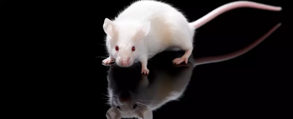
A recent groundbreaking study by researchers at Stanford University has set a new standard in biological imaging by successfully turning parts of living mice see-through. The innovative method involves the use of a biologically-safe dye that alters the light scattering properties of the cells’ surrounding fluids, enabling researchers to observe the intricate workings of an organism’s internal structures while it is alive. This remarkable feat opens up a world of possibilities in various fields, from medical diagnostics to cosmetic procedures.
Traditional imaging techniques often face challenges when it comes to visualizing deep tissues and organs due to light scattering in biological materials. The difference in refractive indices between tissues and fluids causes light to scatter in all directions, resulting in opacity. However, by introducing a dye that selectively absorbs specific wavelengths of light, the researchers were able to modify the refractive index of the surrounding fluid, significantly reducing scattering and rendering the tissues transparent.
The key to achieving transparency lies in the absorption properties of the dye, which absorbs light of particular wavelengths, thereby affecting the refractive index of the fluid. By leveraging the Kramers-Kronig relationship, a mathematical connection between absorption and refraction, the researchers were able to finely tune the dye to optimize transparency while maintaining biocompatibility. This breakthrough not only offers a non-invasive way to visualize internal structures but also highlights the potential for future advancements in biomedical imaging.
The implications of this technology are vast, with potential applications ranging from medical procedures to scientific research. By making veins more visible for blood drawing, simplifying laser-based tattoo removal, and aiding in early cancer detection, this innovative approach could revolutionize the way we study and diagnose diseases. Furthermore, the cost-effectiveness and efficiency of the dye make it a promising tool for widespread use in various fields.
Challenges Ahead
While the results are promising, there are still obstacles to overcome, particularly when applying this method to human subjects. The researchers acknowledge that human skin is significantly thicker than mouse skin, posing challenges in achieving the same level of transparency. However, they are optimistic about the potential for further exploration and refinement of the technique to adapt it for human use.
The see-through mice study represents a major milestone in the field of biological imaging, demonstrating the power of interdisciplinary collaboration and innovation. By harnessing the principles of optics and biology, the researchers have paved the way for a new era of transparent imaging that could revolutionize the way we understand and treat diseases. As the technology continues to evolve, the possibilities for improving healthcare and advancing scientific knowledge are endless.

Leave a Reply